Emerging Tech
A familiar bridge to digital pathology
Digital tools should help pathologists work, not make familiar tasks harder.
A pathologist's hands know a microscope. Moving a slide, finding a small area, changing magnification: these are practiced movements. Going digital should not mean losing that fluency.
I recently met with Dr. Aravindhan Sriharan, founder of PixCell Pathology, for a demonstration of his product. It brought me back to a question that matters as we move from glass slides to digital images: how do we bring people with us?
Digital images, familiar movements
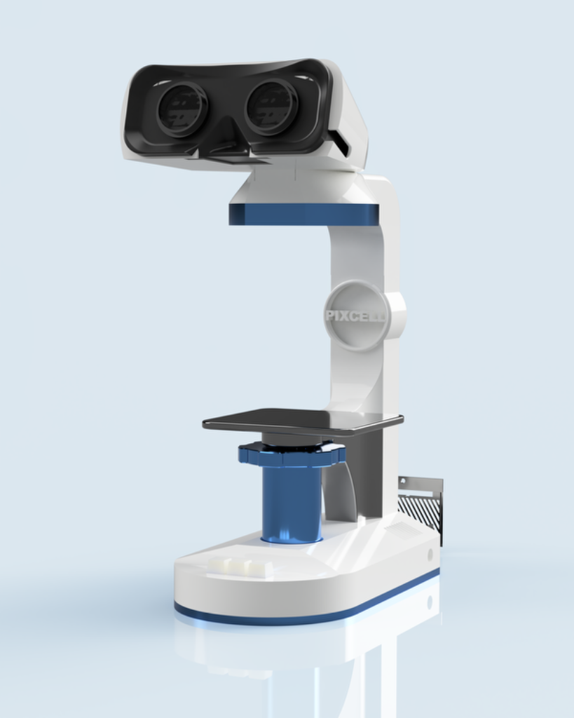
PixCell's SmartScope approaches that question through the interface. It uses a microscope-like viewing system and a movable mock slide to navigate a whole-slide image. Moving the physical slide changes the part of the digital image being viewed. A physical control changes magnification, while a tablet presents a virtual slide tray.
This is a way to view and navigate images that have already been digitized. It does not remove the need to prepare the tissue, make a good slide and scan it.
The idea is a bridge: keep familiar movements while opening the door to digital workflows. That may be more comfortable for some pathologists than navigating with a mouse. It is not a reason to assume one interface will suit everyone. The useful test is whether it helps people do accurate work without adding friction.
What the early evidence shows
A 2023 American Society of Dermatopathology meeting abstract compared two dermatopathologists reviewing 18 routine skin biopsies. Their mean diagnostic times were 117 seconds with mouse-based navigation, 56 seconds with the SmartScope prototype and 24 seconds with a conventional microscope.
In that small study, SmartScope was faster than the mouse-based interface but slower than the microscope. That is an encouraging reason to study the interface, not proof of a general productivity gain. Larger studies across readers, specimen types and real laboratory workflows would help show where it fits.
Two readers, 18 routine skin biopsies. These are study means, not expected times for every pathologist or case. Source: ASDP 2023 abstract.
Beyond the eyepiece
PixCell's website describes a broader set of services around the device. LabLink is presented as a marketplace for pathologists, histotechnologists and laboratory staff, including permanent and temporary work. The company also describes remote access, second-opinion collaboration and opportunities to volunteer expertise in underserved settings.
For researchers, it advertises a software development kit for running models built with PyTorch or TensorFlow, data collaboration and partnerships for clinical validation, hardware testing and grant applications. These are company-described possibilities, not independently verified measures of how widely the services are being used.
The AI App Store needs a careful distinction. Its current page calls the listed algorithms "upcoming." Breast cancer detection, malaria screening and Ki-67 counting are marked as under development. DermPath Assist is marked Research Use Only. A listing is not evidence that a tool is cleared for clinical diagnosis or ready for routine patient care.
The next five to ten years
In February 2026, KLAS estimated that fewer than 15% of US healthcare organizations had selected a digital pathology vendor and were at different stages of deployment and adoption. That does not mean 15% of all pathologists, slides or laboratories are fully digital.
I would like to see adoption reach 85% over the next five to ten years. That is my aspiration, not a forecast. Reaching it would require more than a new viewer: reliable scanning, storage, integration, training, validation and a clear reason for laboratories to make the investment.
AI may strengthen that reason, from helping a pathologist review an image to research on information that could help guide treatment. But diagnostic assistance, prediction of treatment response and selection of treatment are different tasks. Each needs evidence for its intended use. We should not assume that a familiar interface, or an AI marketplace, makes those possibilities clinically established.
The people behind the technology
That thread runs through our earlier digital pathology adoption and careers piece: new images require new workflows and skilled people. Our Before the Slide lung-biopsy piece starts even earlier, with getting useful tissue in the first place. A digital viewer cannot repair a missing or inadequate specimen.
We have talked about the digital pathology coordinators at NYU: people who prepare slides for scanning, check image quality and keep the workflow moving. We have also remembered Freida Carson's lesson: the slides we make determine the slides we scan. Neither foundation disappears when we add a digital viewer. NYU coordinators · Freida Carson
Our earlier Yale example made another distinction: an AI flag is a reason to look again, not a diagnosis. The same discipline belongs here. The pathologist still has to judge the evidence. Read the Yale second-check piece.
SmartScope is one approach worth examining because it starts with the way pathologists already work. Other interfaces deserve the same attention. The goal is not to make the work look more digital. It is to help people do it well.
Dr. Deepak Mohan · Doctor's Doc MD
For education only. This is not personal medical advice or a product recommendation.